
Revolutionary 3D Mapping of the Clitoris Unveiled
In a landmark development for anatomical science, researchers have published the first comprehensive 3D map of the clitoris. This breakthrough comes from Amsterdam University Medical Centre, where scientists used advanced X-ray imaging to scan two female pelvic samples, creating an unprecedented visualization of this crucial organ.
Addressing Historical Knowledge Gaps
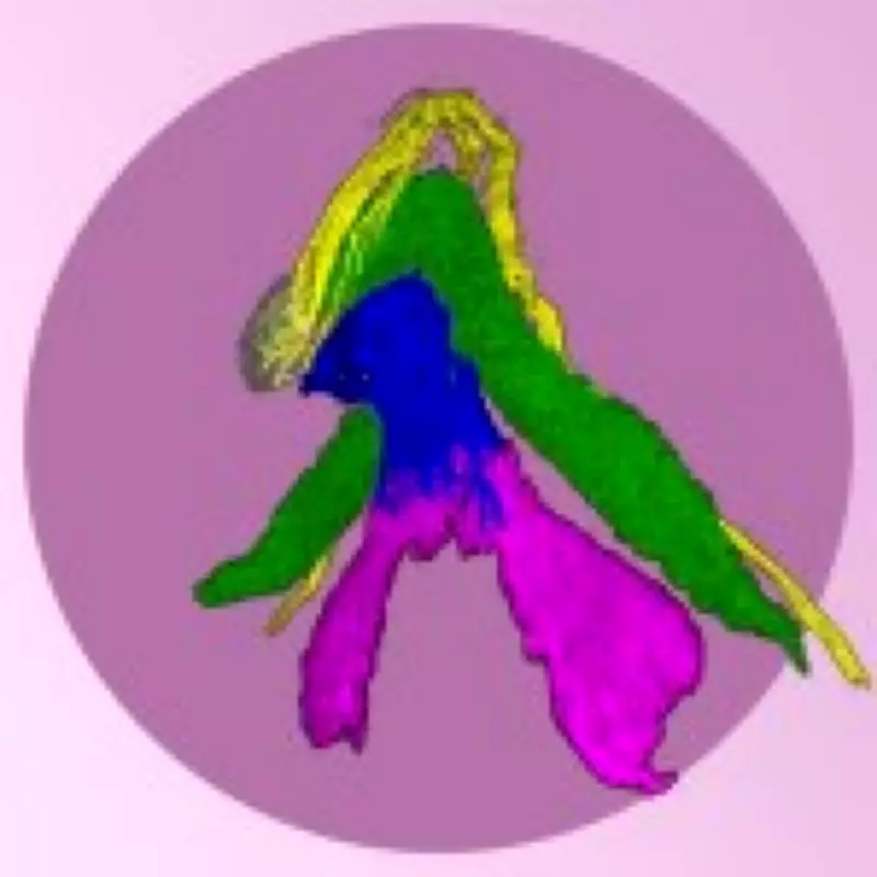
The study, authored by Ju Young Lee, Demi Alblas, Adam Szmul and Daniel Doctor, reveals that the clitoris contains approximately 10,000 nerve endings, confirming its status as the only human organ dedicated solely to pleasure rather than reproduction. The new mapping shows the glans branches out in what researchers describe as a "tree-like" fashion, with five distinct nerve branches ranging from 0.2mm to 0.7mm in width.
This detailed anatomical mapping arrives 28 years after the first 3D map of the penis was created, highlighting the historical disparity in research attention between male and female sexual anatomy. The authors note that cultural taboos around female sexuality have hindered scientific investigation for centuries, with the clitoris being described as the "shameful member" in 16th century France and not appearing in standard anatomy textbooks until the 20th century.

Practical Applications and Educational Impact
Looking forward, the research team hopes their findings will inform various surgical procedures involving the vulva, including those related to childbirth, gender affirmation surgery, and reconstruction surgery. The detailed nerve mapping could help surgeons avoid damaging sensitive structures during operations.
The timing of this discovery coincides with concerning statistics about public knowledge of female anatomy. A recent Lovehoney survey of 2,000 UK adults found that only 3% of women and 2% of men could correctly identify a diagram of the clitoris's internal structure. When shown the image, 24% of participants thought they were looking at a heart, 13% identified it as the vagina, and 10% believed it showed ovaries.
Understanding Clitoral Sensitivity
While 90% of Brits claim to know the location of the glans clitoris (the visible part), only 30% could accurately label it on a diagram, with women performing only marginally better than men (30% versus 29%). This knowledge gap extends to understanding how the clitoris functions during sexual activity.
Sexual health expert Sarah Mulindwa explains that the clitoris's nerve endings aren't evenly distributed, which explains why many women prefer targeted stimulation. "It's very likely that one side, or a particular spot, of the clitoris is going to be much more sensitive and attuned to pleasure than another spot," she notes.
This understanding aligns with findings from sex education site OMGYES, whose survey of 20,000 women revealed that one in eight prefer giving extra attention to specific areas of the clitoris during masturbation, a technique known as "accenting."
Correcting Historical Misconceptions
The study references the fact that when the clitoris was finally included in the 38th edition of Gray's Anatomy in 1995, it was incorrectly described as a "small version of the penis." This new research definitively corrects that misconception while providing the detailed anatomical understanding that has been lacking for centuries.
As the scientific community gains this new understanding of clitoral anatomy, the researchers emphasize that their work represents more than just academic progress—it provides concrete tools for improving medical care and sexual education for people with vulvas worldwide.