
In a groundbreaking advancement for medical science, researchers in Switzerland have successfully 3D printed a human ear that closely mimics the flexibility and mechanical properties of natural tissue. This development marks a significant step forward in the field of biofabrication, potentially revolutionizing reconstructive surgery for individuals with ear deformities or injuries.
From Laboratory to Lifelike: The Evolution of 3D-Printed Ears
For over three decades, scientists have pursued the creation of functional ears from living tissue. In 2016, a team led by ETH professor Marcy Zenobi-Wong gained international recognition for producing a 3D-printed ear, though it lacked the full elasticity of natural ears. Now, a collaborative effort from ETH Zurich, the Friedrich Miescher Institute, and the Cantonal Hospital of Lucerne has achieved a more realistic replica. Using human ear cartilage cells, the team engineered elastic cartilage in the laboratory with mechanical characteristics akin to real tissue.
Overcoming Key Challenges in Tissue Engineering
The research, detailed in the journal Advanced Functional Materials, addresses one of the biggest hurdles in ear reconstruction: elastin, the protein that provides flexibility. Lead author Philipp Fisch, a senior researcher in Zenobi-Wong's Tissue Engineering and Biofabrication Group, emphasized the goal of achieving stability in the lab before implantation. 'We aren't implanting soft tissue in the hope that it remains stable in the body. Instead, we want to achieve that stability in the laboratory,' Fisch explained.

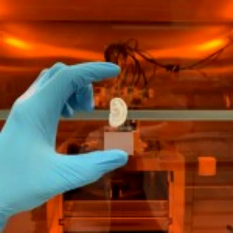
To create the cartilage, cells were extracted from small remnants during routine ear-correction surgeries. These cells were multiplied in a nutrient-rich solution and a controlled culture environment to prevent scar tissue formation. The expanded cells were then embedded in a gel-like 'bioink' and shaped into ears using a 3D printer. After maturation in an incubator for several weeks, the tissue strengthened, with Fisch highlighting four critical factors: optimized cell proliferation, adjusted material properties, increased cell density, and controlled maturation environment.
Implications for Medical Treatment and Future Prospects
This innovation holds promise for people affected by accidents, fires, or congenital conditions like microtia, which impacts approximately four in every 10,000 children. Current reconstruction methods often involve harvesting cartilage from a patient's ribs, leading to pain, scarring, and chest deformities, with results that are typically stiffer than natural ears. In animal tests, the bioengineered ear retained its shape and elasticity after six weeks under rat skin, demonstrating stability similar to natural cartilage.
However, challenges persist. Progress in producing elastic ear cartilage is slow, with experiments taking three to four months as researchers seek the elusive elastin blueprint. Fisch noted, 'We've been working on this problem in our group for over ten years. When it comes to biofabrication of tissue, or tissue engineering as it's also known, swift progress is rare to see.' Interest is growing, with Fisch receiving inquiries from parents of children with microtia shortly after the study's publication.
Looking ahead, Fisch remains cautiously optimistic, aiming to identify the elastin network blueprint within five years. Subsequent clinical studies and regulatory approvals will be necessary before lab-grown ears can be integrated into routine medical practice, offering a less invasive and more effective solution for ear reconstruction.